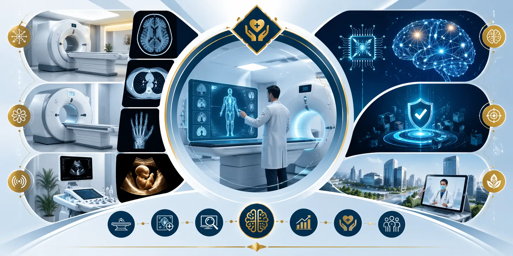
What Is Diag Image Technology?
Definition of Diagnostic Imaging
Diagnostic imaging refers to a suite of non-invasive technologies used to create visual representations of the internal structures and functions of the body. These images help physicians detect abnormalities, monitor disease progression, guide treatment decisions, and assess how well treatments are working — all without surgical intervention.
Today, diagnostic imaging is a core component of healthcare delivery, influencing roughly 70% of clinical decisions across specialties.
Why Diag Images Are Essential in Modern Healthcare
Diag images have become indispensable because they:
- Reveal internal structures invisible to the naked eye.
- Enable early detection of disease, often before symptoms emerge.
- Reduce the need for exploratory surgery.
- Provide objective data for tailored treatment planning.
Clinicians rely on these images to detect fractures, tumors, vascular diseases, neurological conditions, pregnancy abnormalities, and more. In emergency settings, rapid imaging can mean the difference between life and death.
How Diag Image Differs from Traditional Diagnosis
Traditional diagnosis often relied on physical examination, patient history, and basic lab tests. While these remain critical, they can miss critical internal details. Diag imaging introduces a visual dimension — allowing clinicians to see what is happening inside the body in real time and at high resolution.
Evolution of Diagnostic Imaging Systems
Early Medical Imaging Methods
The earliest imaging modality was the X-ray, discovered in 1895. X-rays quickly became a staple for diagnosing bone fractures and chest conditions. Over time, advances in physics, computing, and digital electronics have given rise to more sophisticated techniques.
Transition from Analog to Digital Imaging
The transition from film-based imaging to digital systems vastly improved image quality, storage, and accessibility. Digital imaging allowed clinicians to enhance, zoom, and share images instantaneously.
Today, over 85% of hospitals worldwide use advanced digital imaging technologies that integrate seamlessly with electronic health records (EHRs).
Rise of Advanced Imaging Modalities
Beyond X-rays, modalities such as CT, MRI, and PET scan provide increasing levels of anatomical and functional detail. These technologies have transformed how diseases are identified, staged, and monitored.
AI and Automation in Modern Diag Image Systems
Artificial intelligence (AI) is reshaping diagnostic imaging by improving detection rates, speeding interpretation, reducing errors, and enhancing workflow efficiency. In the coming decade, AI integration in medical imaging is predicted to grow rapidly, helping clinicians interpret images with remarkable precision.
Types of Diag Imaging Modalities Explained
X-rays – Fast and Cost-Effective Imaging
X-ray imaging uses ionizing radiation to capture images of dense tissues like bones. It’s fast, widely available, and relatively inexpensive. Common uses include diagnosing bone fractures, dental issues, chest conditions, and lung infections.
A typical chest X-ray exposes patients to about 0.1 mSv of radiation, which is considered low and generally safe when medically justified.
CT Scans (Computed Tomography)
How CT Scans Work
CT scans use multiple X-ray images from different angles to create cross-sectional views of the body. These high-resolution images help visualize soft tissues, organs, blood vessels, and skeletal structures with greater detail than simple X-rays.
Common Clinical Applications
CT scans are crucial in diagnosing complex conditions such as:
- Traumatic injuries
- Tumors
- Cardiovascular disease
- Pulmonary conditions
However, because they use ionizing radiation, clinicians balance benefits against radiation exposure risks. One study suggests CT scans may contribute to up to 5% of cancer cases annually — underscoring the importance of judicious use.
MRI (Magnetic Resonance Imaging)
MRI uses magnetic fields and radio waves to create detailed images of soft tissues like the brain, spinal cord, joints, and organs — all without ionizing radiation.
MRI is especially valuable for:
- Neurological conditions
- Musculoskeletal injuries
- Tumor characterization
MRI usage has increased about 50% globally in the last decade, reflecting its diagnostic importance.
Ultrasound Imaging
Ultrasound uses high-frequency sound waves to capture real-time images of internal structures. It’s widely used in:
- Obstetrics (e.g., monitoring pregnancy)
- Abdominal organ assessment
- Cardiac evaluation
Ultrasound is safe, non-invasive, and free of radiation, making it the modality of choice for many patients.
PET Scans (Positron Emission Tomography)
PET scans involve injecting a small amount of radioactive material that accumulates in metabolically active tissues. It’s especially useful for cancer detection, staging, and monitoring treatment response.
How Diag Imaging Improves Early and Accurate Diagnosis
Precision Meets Speed in Emergency Care
In emergencies such as strokes or traumas, time is critical. Rapid imaging can detect brain bleeds, fractures, or vascular blockages within minutes, allowing clinicians to initiate treatment without delay.
Early Disease Detection and Risk Reduction
Diag imaging can identify disease in its pre-symptomatic stages, speeding interventions that save lives. For example, mammography detects early breast cancers with sensitivity often above 85%, making it a key screening test for women.
Minimally Invasive but Deeply Insightful Diagnostics
Before these technologies, diagnosing internal conditions often required exploratory surgery or invasive procedures. Today, imaging reveals internal pathology without physical intrusion — reducing patient risk and recovery time.
Reducing Diagnostic Errors with Imaging Support
Diagnostic errors occur in a significant portion of clinical cases, contributing to adverse outcomes. AI and advanced diag image systems can reduce errors by up to 30–40% by supporting radiologists and clinicians with enhanced pattern detection.
Advanced Visualization and Measurement Tools in Diag Image
Image Processing and Enhancement Tools
Modern diag image systems include tools for adjusting contrast, magnification, and resolution — enabling clinicians to see subtle features that could signal disease.
Measurement and Goniometry Capabilities
These tools help quantify distances, angles, and areas on images — important in fields like orthopedics, cardiology, and oncology.
3D Imaging and Reconstruction
3D reconstruction allows clinicians to rotate and visualize body structures in three dimensions — a powerful tool for surgical planning and assessment of complex conditions.
Automated Display and Hanging Protocols
Automated hanging protocols organize images on screen in clinically logical layouts, speeding interpretation and minimizing errors.
Image Comparison and Longitudinal Analysis
Clinicians can compare current diag images with previous ones to track disease progression or healing — a vital feature for chronic disease management.
Technology Powering Modern Diag Image Systems
Digital Imaging Infrastructure
Digital systems store images electronically, allowing easy sharing across departments and with patients via portals. This digital transformation has made medical imaging faster, more accurate, and more integrated with patient care.
AI-Powered Diagnostic Imaging
AI algorithms analyze images faster and often more accurately than traditional methods. Advanced AI systems have achieved diagnostic accuracy rates exceeding 90–95% in detecting certain conditions, and can lower false positive or false negative rates compared to human interpretations.
These tools do not replace clinicians — they augment decision-making by highlighting abnormalities and quantifying regions of interest.
Integration with PACS and EHR Systems
Picture Archiving and Communication Systems (PACS) and EHR integration ensures that diag images and reports are stored centrally, securely, and accessible whenever needed. This interoperability streamlines clinical workflows.
Cloud-Based Image Storage and Access
Cloud platforms enable remote access to medical images, supporting telemedicine, multi-center collaboration, and expedited care delivery.
Role of Diag Image Centres in Healthcare Delivery
What Happens at a Diagnostic Imaging Centre
Patients typically:
- Check in and provide medical history.
- Get instructions from technicians or radiologists.
- Undergo imaging procedure based on clinical need.
- Have results processed and shared with their primary doctor, often within hours.
Benefits of Specialized Imaging Centres
Imaging centres often provide:
- Faster appointments than hospitals
- Specialized protocols
- Advanced equipment
- Dedicated radiology expertise
Faster Access, Lower Costs, and Better Outcomes
Centres often deliver services more efficiently and at lower cost than larger hospital systems — improving access for patients.
Patient Experience and Online Image Portals
Many imaging centres now offer online portals where patients can review their images and share them with multiple care providers.
Applications of Diag Images Across Medical Specialties
Oncology – Cancer Detection and Monitoring
Diag imaging helps:
- Detect tumors early
- Monitor response to treatment
- Guide biopsies and radiation therapy
PET-CT, MRI, and contrast-enhanced CT scans are indispensable tools in oncology.
Cardiology – Heart Imaging and Risk Assessment
CT angiography and echocardiography visualize coronary arteries, structural heart conditions, and cardiodynamics, enabling personalized treatment planning.
Neurology – Brain and Spinal Cord Imaging
Orthopedics – Bone, Joint, and Muscle Imaging
MRI and CT scans provide detailed views of joint injuries, fractures, and degenerative diseases.
Emergency Medicine and Trauma Care
Rapid diag imaging identifies life-threatening conditions such as internal bleeding, fractures, and stroke — saving crucial time in emergencies.
Pediatrics and Preventive Screening
Safe imaging modalities like ultrasound are widely used in children to monitor growth and diagnose congenital anomalies.
Importance of Diagnostic Accuracy in Healthcare
Impact of Diagnostic Errors on Patient Outcomes
Diagnostic errors contribute significantly to adverse outcomes and malpractice claims, affecting up to 15% of healthcare cases.
Cost of Misdiagnosis to Healthcare Systems
Incorrect diagnoses lead to unnecessary tests, delayed treatment, increased hospital stays, and higher costs, underscoring the need for improved imaging precision.
How Diag Image Improves Clinical Confidence
Advanced imaging increases confidence in clinical decisions, reducing unnecessary interventions and improving patient outcomes.
Decision Support and Quantitative Analysis Tools
Quantitative tools help clinicians measure and track changes over time, increasing diagnostic rigour.
Benefits of Diag Image Technology
Improved Diagnostic Confidence
Diag images help reduce guesswork and provide clarity in complex cases.
Enhanced Clinical Workflow Efficiency
Digital and AI tools speed up reporting times, standardize workflows, and reduce interpretation burden.
Non-Invasive and Patient-Friendly Diagnosis
Unlike surgical exploration, diag imaging is non-invasive and often painless.
Better Treatment Planning and Monitoring
Image-based assessments allow tailored treatment plans and continuous monitoring.
Challenges and Limitations of Diag Image Systems
Cost and Insurance Coverage Issues
Specialized scans like MRI or PET can be expensive — sometimes limiting access for patients without comprehensive insurance.
Radiation Exposure and Safety Concerns
Techniques like CT and X-ray involve ionizing radiation, necessitating careful justification and dose optimization.
AI Bias and Data Quality Challenges
AI systems must be trained on diverse datasets to ensure equitable performance and avoid interpretation bias.
Accessibility in Remote and Underserved Areas
Access to advanced imaging technologies remains uneven, especially in low-resource regions where trained personnel and equipment may be scarce.
Data Security and Privacy Risks
Storing and transmitting medical images requires strict compliance with privacy standards to safeguard patient data.
Future of Diag Image Technology
AI-Driven Predictive Diagnostics
AI will continue to elevate diagnostic capabilities, potentially predicting disease progression and outcomes based on imaging patterns.
Augmented Reality and Imaging Visualization
Emerging tools may allow clinicians to overlay imaging data directly onto patients during procedures, enhancing precision.
3D Printing from Diagnostic Images
Surgeons can use 3D printed models of patient anatomy for pre-surgical planning and simulation.
Personalized Medicine and Imaging Data
Imaging combined with genetic and clinical data will support truly individualized treatment pathways.
Remote Diagnostics and Tele-Imaging
High-resolution imaging, streamed securely, can empower specialists to assess patients worldwide.
Diag Image vs Traditional Diagnostic Methods
Accuracy Comparison
Advanced imaging often detects conditions earlier and more precisely than traditional methods.
Speed and Efficiency
Digital imaging combined with AI dramatically reduces time from capture to diagnosis.
Risk and Patient Comfort
Non-invasive methods reduce physical stress and recovery time.
Cost and Long-Term Value
Although some imaging modalities are costly, early diagnosis often yields long-term savings by preventing advanced disease.
Conclusion: Why Diag Image Is the Backbone of Modern Medicine
Diag image technology is foundational to modern healthcare. It enhances diagnostic accuracy, expedites clinical decisions, reduces invasive procedures, and supports personalized patient care. With continuous innovations — especially in AI and digital integration — the future of diag imaging promises even greater accuracy, accessibility, and clinical impact.
Frequently Asked Questions (FAQs)
What is Diag Image used for?
Diag images capture internal body structures to guide clinicians in diagnosing, monitoring, and treating disease.
Is Diag Image safe for patients?
Many modalities like ultrasound and MRI have no radiation. Others like CT and X-ray use ionizing radiation but are generally safe when medically justified.
How does AI improve Diag Image accuracy?
AI assists clinicians by highlighting abnormalities, quantifying image features, and reducing interpretation time.
Can Diag Image reduce diagnostic errors?
Yes — especially when combined with decision support and AI — which has been shown to significantly reduce interpretation errors.
Is Diag Image compatible with PACS systems?
Yes. Most modern imaging systems integrate with PACS and EHR platforms.
What training is required for Diag Image systems?
Technicians and radiologists undergo specialized training to operate equipment and interpret images accurately.
Read More: Finnorth Explained